
Grzegorz Piotr Waranecki-Eye Disorders-(Poland)-Posted on Dec.16,2014
 Name: Grzegorz Piotr Waranecki
Sex: Male
Age: 43
Nationality:Poland
Diagnosis:1. Optic Nerve Atrophy 2. Glaucoma
Admission Date: 2014-11-17
Treatment time:21 days
Before treatment:
Due to glaucoma and vision deterioration,Waranecki had eyes examination in ophthalmology department 5 years ago. It was detected that his bilateral retinal nerve fiber layers were thinner than normal and optic nerve atrophied. Then he had examination every half a year and found it was progressively thinning. Electronic vision detection showed that it was a progressive constriction of his bilateral upper vision field. He didn’t take any special treatment, and his vision constriction accelerated from the last 6 months.
Since the onset of the disease,the patient’s mentality has been good,normal weight,diet,sleep,defecation and urination.
Admission PE:
Bp: 125/89mmHg; Hr: 70/min, Br: 18/min. Height: 178cm, weight: 75Kg. Waranecki’s nutrition status is normal. His skin and mucous was normal with no yellow stains or petechia. The thorax was symmetrical. The respiratory sounds of both lungs were clear, no rales. The heart sounds were strong, the rhythm was regular, and there was no obvious murmur in the valve area. The abdomen was flat and soft with no obvious masses. Through palpation, the liver and spleen were not enlarged. There was no edema of both lower limbs.
Nervous System Examination:
Waranecki was alert and his speech was clear. His memory, orientation and calculation ability were normal. Both pupils were equal in size and round, the diameter was 2.5mms. Both his eyes were sensitive to direct light reflex and consensual reflex. Color vision was normal. Corrected vision of left and right eye is 0.6 and 0.8 for 3 meters visual chart. Both eyeballs could move freely to each side. There was no nystagmus in both eyes. Under ophthalmoscope: both fundus were presented with jacinth. Equator area had little dark sediment and eyecup enlarged. Boundary of macular area was unclear. Retinal diopter was extremely low. The A/V ratio was 1:3. The forehead wrinkle pattern was symmetrical, the nasolabial sulcus was equal in depth, the teeth were symmetrical and the tongue was centred in the oral cavity. There was flexible movement in the neck. The muscle tone of all four limbs was normal; the muscle strength of all four limbs was at level 5. The abdominal reflexes were normal. The tendon reflexes of the four limbs were normal. The Pathological signs were negative. The deep sensation and superficial sensation were normal. The coordinated movements were normal. There was no meningeal irritation.
Retinal Nerve Fiber Layer Examination:Left 0.14mm2,right 0.11mm2 (0.88-1.87).
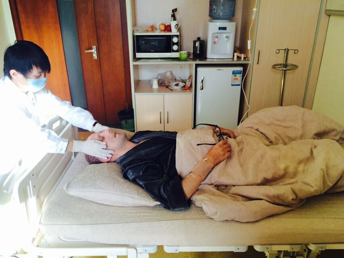
Treatment:
We gave Waranecki a complete examination and he was diagnosed with Optic Nerve Atrophy and Glaucoma. He received treatment to control IOP, activate or launch his neuro recovery and nerve regeneration and activate his own stem cells. He also received treatment to improve the blood circulation in order to increase the blood supply to the damaged nerves, nourish the neurons and adjust his immunity.
Post-treatment:
Both visions have improved obviously. Corrected vision of left and right eye is 1.0 and 1.2 for 3 meters visual chart. Under ophthalmoscope: both fundus are presented with jacinth. Part of dark sediment of equator reduced. The border of macular area is clearer than before. Retinal diopter increased 30%. The A/V ratio is 1:3 and it is nearly normal.
(Download the Windows Media Player Firefox Plugin if you are using Firefox browser.
To know more,Please read Using the Windows Media Player plugin with Firefox.)