
Antonio Romani-Retinitis pigmentosa-(America)-Posted on Feb.2nd, 2015
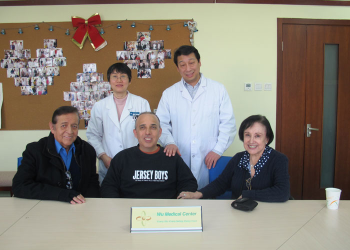
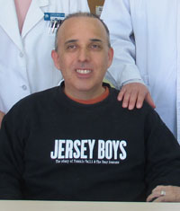 Name: Antonio Romani
Sex: Male
Country: America
Age: 47 years
Diagnosis: Retinitis pigmentosa
Date: Jan. 6, 2015
Days Admitted to Hospital: 23 days
Before treatment:
Antonio Romani was presented with left red eye for about 20 years ago, with no visible diminution of vision. The ophthalmologist diagnosed him with retinitis pigmentosa through various examinations. He began to take Vitamin A 15000iu/ Qn. Then he had a progressive deterioration of his vision in both eyes. It aggravated day by day. His day vision and peripheral visual field was also defected. It was difficult for him to see objects during night 7 years ago. He couldn’t read or drive during day time 5 years ago. His vision decreased fast in 3 years, he couldn’t distinguish between colors, he could only see the outline of bigger objects and fingers in 50cm, but he couldn’t distinguish the number of fingers. He wants to have a better treatment, so he came to our hospital.
He was in good spirit. His weight was stable. His diet, sleeping, urination and excrement were normal.
Admission PE:
Bp: 127/81mmHg; Hr: 83/min. He’s growth and nutrition was normal; his skin mucosa was without hemorrhaging spots or yellow stains. His thorax was symmetrical. The respiration of both lungs was clear, without moist rales. The heart sound was strong, and the cardiac rhythm was regular, and there was no obvious murmur in the valves. The abdomen was flat and soft. The liver and spleen were normal. Both lower limbs were not dropsically.
Nervous System Examination:
He was alert and his speech was clear. His memory, orientation and calculation ability were normal. The diameter of both pupils was 3.0mms and both pupils were sensitive to direct and indirect light reflex. His peripheral visual field was defected; central field of vision was blurry. He could see the outline of bigger object from 1.5 meters distance, and he could also see the finger shake but could not distinguish the number form 0.5 meter distance. He couldn’t distinguish colors. Through use of an ophthalmoscope: there was no extravasation on the fundus. The boundary of optic papilla was clear no dropsy. The color was a little white. The color of retina was light yellow. AV ratio of both eyes was 1:3. The boundary of macula flava retinae was not clear. There were a lot of black osteoid lees on the ambitus. The movement of eyeballs to each side was good and there was no obvious nystagmus. The forehead wrinkle pattern was symmetrical. The nasolabial sulcus was equal in depth. The tongue was centered. There was free movement in the neck. The muscle tone of all four limbs was almost normal; the muscle strength of all four limbs was almost at level 5.The tendon reflexes in four limbs were normal. The pathologic reflex was negative. The deep and shallow sensation was normal. The coordinated movements were normal.
Treatment:
We gave him a complete examination and he was diagnosed with retinitis pigmentosa. He received treatment to repair and revive the retina and optic nerves, activated his stem cells, nourish the neurons, dilate the blood vessels, and regulate the immune system.
Post-treatment:
After the 3 weeks of treatment, his vision was better. He could distinguish fingers and some colors’ shades; sometimes he could distinguish black, pink and gray. He can also distinguish bigger objects’ shape from 1.5-2 meters.Through use of an ophthalmoscope: the color of retina is better; AV ratio 2:3, the black osteoid cells on the ambitus was less, especially on the yellow spot part. At present, Antonio Romani’s treatment has been completed and the requirements for discharge have been met.
(Download the Windows Media Player Firefox Plugin if you are using Firefox browser.
To know more,Please read Using the Windows Media Player plugin with Firefox.)