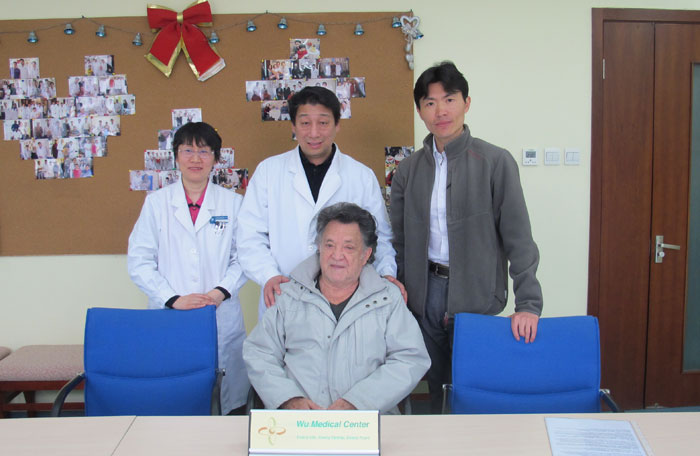
Angel A Cueva-Diabetic Retinopathy-(Ecuador)-Posted on Apr.7th, 2015
 Name: Angel A Cueva
Sex: Male
Country: Ecuador
Age: 75 years
Diagnosis: Type2 Diabetes, Diabetic Retinopathy, Hypertension level 3(Very high-risk), Cataract
Date: Mar. 14th, 2015
Days Admitted to Hospital: 22 days
Before treatment:
The patient experienced retinal hemorrhage about 15 years ago. He had ocular hypertension and hypertension. He was treated with laser surgery to stop the bleeding. After the therapy, his vision gradually decreased because he bled several times from the fundus. He had 3 times laser surgery in 15 years but his vision had no improvement. His night vision was very bad as well as his day vision. He could hardly read especially with right eye. He could only see fingers from the distance of 5cm. His left eye could see big objects from the distance of 5 meters. He wants to have a better treatment, so he came to our hospital.
He was in good spirit. His weight, eating, urination and excrement were good. He couldn’t sleep well and had astriction.
Admission PE:
Bp: 101/67mmHg, Hr: 67/min, Br: 19/min. body temperature: 36.5 degrees. He’s growth and nutrition were normal. His skin had normal mucosa without petechia or stained yellow. His thorax was symmetrical. The respiration of both lungs was clear, without moist rales. The heart sound was strong and the cardiac rhythm was regular and there was no obvious murmur in the valves. The abdomen bulged and soft, with no masses. The liver and spleen were normal. His blood sugar:13-22mol/L, blood urea nitrogen: 10.49mmol/L.
Nervous System Examination:
He was alert and his speech was clear. His memory, orientation and calculation ability were normal. The diameter of left pupil was 2.0mms and the shape was irregular. The diameter of right pupil was 2.0mms and it was equal in circumference. Both pupils were not sensitive to light reflex. The right eye lost central field of vision, only had peripheral visual field and the vision was not very good. The left eye could only see the number of fingers at the distance of 5cm from temporal side. The visual field of right eye was normal, 3-meter visual acuity test chart: 0.12. The movement of eyeballs to each side was good and there was no obvious nystagmus. The bottoms of eyes examination: left side: the color of the bottoms of eyes was partly light yellow other parts were somehow orange. AV ratio: 1:4. The boundary of macular area was not clear. The optic papilla was normal without exudation. Right side: the color of the bottoms of eyes was light yellow. AV ratio: 1:4. The macular area was depauperated and dilacerated. The boundary of optic papilla was clear. There was some exudation around the veins. Both sides of the bottoms of eyes had level 3 arteriosclerosis. The forehead wrinkle pattern was symmetrical. The nasolabial sulcus was equal in depth. The teeth were symmetrical and the tongue was centred. There was free movement in the neck. The muscle tone of all four limbs was almost normal. The muscle strength of all four limbs was almost at level 5. The abdominal reflex was weak. The reflex of both side biceps brachii and triceps brachii were normal. The patella tendon reflex and achilles reflex were lower than normal. Both side Hoffmann’s sign was negative, the pathologic reflex of left lower limb was positive, the pathologic reflex of left lower limb was negative. The deep and shallow sensation was normal. The coordinated movements were normal. The meningeal irritation sign was negative.
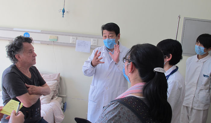
Treatment:
We gave him a complete examination and he was diagnosed with type2 diabetes, diabetic retinopathy, hypertension level 3(very high-risk) and cataract. He received treatment to widen blood vessels, improve the blood circulation, nourish nerves and lower the blood pressure and blood sugar. He also had rehabilitation exercise.
Post-treatment:
After the treatment, his blood pressure and blood sugar were controlled better. Blood pressure: 110-135/65-85mmHg. He used less insulin (the dose decreased from 66iu to 58iu). His blood sugar controlled better, fasting blood sugar: 3.7-6.4mmol/L, 2 hours after meal blood sugar: 7.0-12.0mmol/L. the renal function urea nitrogen decreased from 10.49mmol/L to 8.60mmol/L. His right eye could see fingers at the distance of 0.5 meter but couldn’t distinguish colors. His left eye could see big object more clearly from the distance of 5 meters. Through ophthalmoscope: left side: the bottoms of eyes was orange, optic papilla was light yellow, the boundary of optic papilla was clear without dropsy. The boundary of macular area was clear. The blood circulation was better than before. AV ratio was 1:3. There was no exudation around the veins. Right side: the bottoms of eye was light yellow, the boundary of optic papilla was clear. The macular area was depauperated and dilacerated, AV ratio was 1:3. There was some exudation around the veins. Both sides of the bottoms of eyes had level 3 arteriosclerosis.