Lucca Exequie Manrique Ortiz-Binocular retinal detachment-(Argentina)
 Patient name: Lucca Exequie Manrique Ortiz
Gender: Male
Age: 6 years old
Nationality: Argentina
Diagnosis: Binocular retinal detachment
Before treatment:
Vision loss in both eyes and no sense of light for more than 6 years, left pupil atrophy.
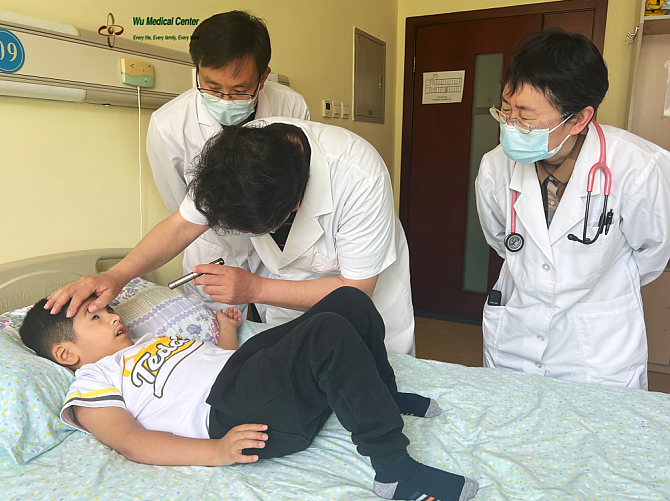
Physical examination on admission:
Blood pressure 80/54mmHg, heart rate 98 beats/min, breathing 24 breaths/min, body temperature 36 degrees, height 71cm, weight 24Kg, good nutrition, no damage to the skin and mucous membranes of the whole body, no cyanosis of the lips, no redness and swelling in the oropharynx. Clear breath sounds in both lungs. There was no bulge in the precordial area, the heart sounds were strong and uniform, and there were no murmurs in each membranous area. The abdomen was flat and soft, and the liver and spleen were not enlarged. Physiologic curvature of the spine was present, and there was no edema in both lower extremities.
Neurological examination:
Mental clarity, fluent language, basically normal calculation ability and orientation ability. The left pupil was atrophied, the right pupil was cloud-like turbidity deposited, about 3 mm in diameter, there was no light perception in both eyes, and the movement of both eyes in all directions was inflexible. The bilateral frontal lines were symmetrical. The tongue was centered when the tongue is extended. The neck had good range of motion, normal muscle tone of the limbs, grade 5 muscle strength of the limbs, and negative pathological signs of the limbs. Superficial sensation was present, coordinate movement was normal, and meningeal irritation signs were negative.
Fundus examination:
The left side could not be peeped in; Right retina funnel-shaped, vitreous fibrosis, fundus pale, poor blood supply.
Treatment process:
After admission, it was clearly diagnosed as "binocular retinal detachment", and mesenchymal stem cells and neural stem cells were given to initiate the development of retina and optic nerve, nourish retinal and optic nerve, improve circulation, regulate immunity, and comprehensive visual rehabilitation and other treatments.
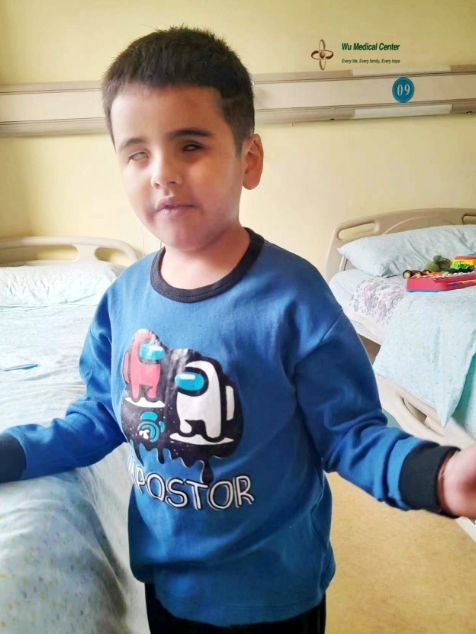
After treatment:
The sensitivity of the patient's right eye to light was improved, the cloud-like turbidity deposition of the right pupil was reduced, the range of motion of the right eye was more flexible than that of admission, and light perception appears, which could track the light source and locate the position of the light source.
Fundus examination:
The fundus of the right eye is light orange-red, the abnormal hyperplasia of blood vessels is reduced, blood supply is improved, and the degree of vitreous fibrosis is reduced.