Maria Cristina-Optic nerve and retinal injury; Glaucoma-(Argentine)
 Patient Name: Maria Cristina
Gender: Female
Age: 75 years old
Nationality: Argentine
Diagnosis: Optic nerve and retinal injury; Glaucoma
Pre‑treatment:
The patient was admitted to hospital with a diagnosis of “optic nerve and retinal injury, glaucoma”, presenting with “progressive bilateral visual loss for more than 5 years secondary to glaucoma”. She developed elevated intraocular pressure 5 years ago and was diagnosed with glaucoma at a local hospital. Her vision deteriorated progressively thereafter. At presentation, the right eye only retained light perception, while the left eye could only see blurry objects at close range with poor object recognition. The right eye had a history of corneal ulcer and retinal detachment. A disc‑shaped silicone sheet had been implanted to regulate intraocular pressure. Nevertheless, visual loss kept progressing. At the time of admission, she was using Dorzolamide 2% / Timolol eye drops (Glaucotensil), Latanoprost 0.005% eye drops (LOUTEN), and Sodium Carboxymethylcellulose 0.5% eye drops (Carboximetilcelulsa Sodica 0.5%). She had a 12‑year history of hypertension, treated with oral Amlodipine 5 mg / Valsartan 160 mg once daily.
Admission physical examination:
Blood pressure: 155/93 mmHg; heart rate: 48‑55 beats per minute; respiratory rate: 20 breaths per minute. The patient had normal physical development and fair nutritional status. No conjunctival congestion or corneal ulcer was noted in both eyes. Breath sounds were clear in both lungs, with no rales detected. Heart sounds were slightly weak with regular rhythm; no obvious murmurs were heard over each valve area. The abdomen was flat and soft; liver and spleen were not palpable.
Neurological examination:
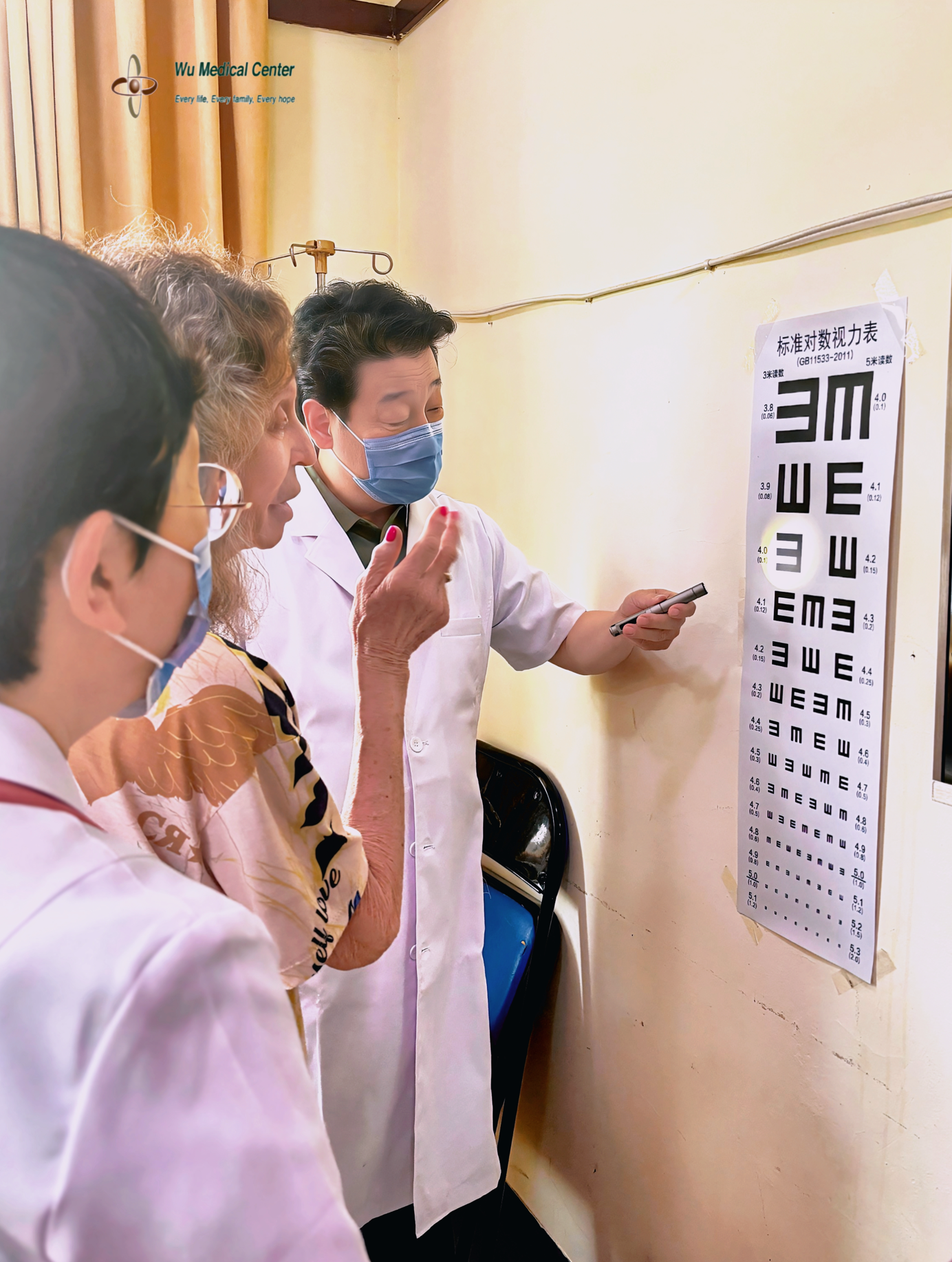
The patient was conscious with fair mental status. Calculation, memory and orientation were basically intact. Bilateral pupils measured 4.0 mm. Direct and indirect pupillary light reflexes were present in the left eye; direct light reflex of the right eye was sluggish, and its indirect light reflex was barely detectable. At 40 cm in front of the standard visual acuity chart, both eyes could vaguely recognize the third positive line (equivalent to 0.1 at 3‑meter reading), corresponding to decimal visual acuity of 0.01. The right eye only had light perception without light localization. Ocular movements were full in all directions for both eyes. Fundus findings for the left eye: no optic disc oedema, ill‑defined disc margin, pale‑yellow disc colour, present fovea centralis with poor foveal reflex, arteriovenous ratio 1:4. Fundus findings for the right eye: no optic disc oedema, relatively blurred disc margin, pale optic disc, fovea centralis nearly invisible, arteriovenous ratio 1:4. Slight pupillary opacification was seen bilaterally. Intraocular pressure was elevated. Forehead wrinkles were symmetrical; tongue protrusion was midline. Muscle strength and muscle tone of neck and four limbs were normal. Tendon reflexes were normal; pathological reflexes were negative. Superficial and deep sensations were preserved; coordination was normal.
Treatment course:
With confirmed admission diagnosis, the patient received CAST therapy plus combined therapy of neural stem cells and mesenchymal stem cells, aiming to repair optic‑nerve and retinal lesions, restore optic‑nerve conduction, nourish the optic nerve and retina, improve internal bodily environment, modulate immunity, together with comprehensive rehabilitation training.
Post‑treatment:
The patient’s visual function is improved: the appearance of bilateral pupils becomes clearer, nearly matching normal eyes. The left eye acquires rudimentary object‑recognition capacity together with partial colour perception: simple objects can be distinguished at 50 cm, finger counting can be performed accurately at about 115 cm, object colours can be correctly identified, and posters can occasionally be seen. At 115 cm in front of the standard visual acuity chart, the patient can recognize the third positive line (equivalent to 0.1 at 3‑meter reading), corresponding to decimal visual acuity of 0.03. For the right eye, light can be detected and precisely localized at 30 cm on the temporal side. Fundoscopic examination shows improvement compared with admission: fundus colour is changed from pale white on admission to light red; the bilateral arteriovenous ratio is improved from 1:4 at baseline to 1:2.